12 setembro 2008

Para ler com muita atenção

D.R. N.º 41 - 27/02/08 ASSEMBLEIA DA REPÚBLICALEI Nº 12-A/2008ASS: Estabelece os regimes de vinculação, de carreiras e de remunerações dos trabalhadores que exercem funções públicas.
Aqui fica a circular do Sindite que ajuda a descodificar o anterior.
Muitas novas regras que vão afectar todos nós...muita atenção, mesmo.

Técnicos de Diagnóstico e Terapêutica passar a Técnicos Superiores de Saúde?

Ministério esgota prazo - Técnicos de Saúde sem propostas

São cerca de dez mil – radiologistas, técnicos de diagnóstico, fisioterapeutas, farmacêuticos, psicólogos, terapeutas. Integram duas carreiras da Função Pública (Técnico Superior de Saúde e Técnicos de Diagnóstico e Terapêutica) mas, ao contrário dos Médicos e Enfermeiros, ainda não receberam qualquer proposta por parte do Ministério da Saúde de revisão das carreiras.
A nova legislação da Função Pública, de Fevereiro, estabelecia um prazo – limite para cada ministério negociar as carreiras especiais, que terminou em Agosto. Contudo, os sindicatos continuam à espera.Edgar Loureiro, do Sindicato dos Técnicos Superiores de Diagnóstico e Terapêutica, acusa o Governo de estar a atrasar este processo para não gastar dinheiro. "O tempo está a correr contra nós, porque o Orçamento de Estado será aprovado em Outubro", refere. E explica que, no caso destes profissionais, a formação é de nível superior, mas os salários não, já que desde 2000 que a carreira está para ser revista.

Correio da Manhã,
05-09-2008
R.A.


É bom ir estando a par destas novidades...que não surgem.

Mobilizar para uma greve a sério?...se calhar é pedir muito.

Link: http://www.sindite.pt/noticia.asp?cod=3

11 setembro 2008

Colapsoterapia com Bolas de Lucite – Imagens de uma técnica do passado

Um artigo meu publicado no Boletim do Hospital Pulido Valente.
Colapsoterapia com Bolas de Lucite – Imagens de uma técnica do passado, apresenta um caso que resulta de uma técnica que já não se realiza há quase 50 anos, muito usada no tratamento da TP.


É um daqueles casos que já não vamos ver muito.


Como está em pdf, tem que ser através do megaupload...disponível para quem quiser.


P.S.: lembrem-se de respeitar os Direitos de Autor.

Saved by Cancer

É um título bizarro para um caso também ele bizarro.

O caso é de um paciente com um Linfoma já conhecido que apresenta, nos exames anteriores, uma Aorta Normal.

O exame realizado agora mostra-no isto...


O Linfoma aumentou consideravelmente e a Aorta apresenta agora um aspecto que pode indicar Dissecção, a formação de um pequeno Pseudoaneurisma, Ulceração ou mesmo um Processo Infeccioso envolvendo a Aorta.

Certo mesmo é o facto de ser a enorme massa envolvente o único factor de suporte e tamponamento que impede esta Aorta de sangrar.
O autor deste artigo inicia-o com o ditado: "O inimigo do meu inimigo, meu amigo é", e provavelmente, nenhum outro se aplicaria tão bem como neste caso.

Link:http://doctordalai.blogspot.com/2008/08/saved-by-cancer.html

05 agosto 2008

Processo de Bolonha - Parecer final

Finalmente uma decisão...

Afinal vai tudo ficar mesmo assim como está...uns licenciados de 3 anos, outros de 4.

Cabe às diferentes escolas decidir se fazem Licenciaturas de 3 ou 4 anos, sendo que, caso se decidam por 4 anos, o estágio profissional terá obrigatóriamente de estar incluido.

Uma decisão que acaba por agradar a todos...hehe

...a mim não...é pena que não nos consigamos entender porque acho que os cursos deveriam ser semelhantes, independentemente da Escola onde são leccionados...mas isso nunca acontece, não é?

As diferenças depois veêm-se cá fora...não vale de nada, mas dá para notar bem.

04 agosto 2008

É esperar que há-de chegar cá...

Um excerto de um artigo de um Blog - Americans for Responsible Imaging que transcreve uma resolução prestes a ser aprovada que autoriza Clínicos de especialidades que não Radiologia a Protocolar e Analisar Exames Radiológicos, sem qualquer intervenção por parte dos Radiologistas.
Só nos USA...apetece dizer...o pior é que, vai a ver, qualquer dia temos uma cópia disto cá...é só algum iluminado lembrar-se disso...hehe
"Fairness" in Medical Imaging Interpretation
A Resolution from the American Misbegotten Association

Resolution 208, Fairness in Medical Imaging Interpretation, is to come before the AMA's House of Delegates shortly, and it is expected to pass. It is introduced by some of our very good friends:


American Society of Neuroimaging

American Association of Neurological Surgeons

Congress of Neurological Surgeons

American Medical Group Association

American College of Cardiology

American College of Gastroenterology

American Gastroenterological Association

American Society for Gastrointestinal Endoscopy

(Associações de várias especialidades unidas por uma causa? Qual $erá a razão?)

So, what do these illustrious physicians call "fair"? Here's the text of the bill and relevant AMA policy:
Whereas, Expenditures for advanced medical imaging services, such as CT, MRI and PET, have significantly increased in this decade, raising legitimate concerns over utilization rates; and
Whereas, These concerns have led payers, state legislatures, government agencies and radiology management companies to consider eliminating reimbursement for in-office imaging and image interpretation by non-radiologists without consideration of their training and certified competence to provide these services; and
Whereas, Available data do not suggest that increased imaging costs are necessarily attributable to in-office imaging services by most medical specialties involved in medical imaging and interpretation; therefore be it
RESOLVED, That our American Medical Association encourage and support the in-office utilization, medical direction and supervision of advanced imaging services by qualified or certified physicians whose utilization of these modalities is within the scope of their specialty practice in accordance with appropriateness guidelines, practice guidelines, technical standards and accreditation standards for the imaging modalities utilized as defined by their specific medical society (New HOD Policy); and be it further
RESOLVED, That our AMA actively oppose efforts by federal and state legislators, regulatory bodies, private payers, public payers and radiology business management companies to restrict the application of advanced imaging services for the diagnosis and treatment of patients when such services are provided as defined by specialty specific appropriateness guidelines, practice guidelines and technical standards for the imaging modalities utilized.
Comentário do Blogger...
Excuse me, but what a bunch of CRAP! It has been proven time and time again that self-referral DOES show increased utilization. Beyond that, what our friends are asking for is a complete disregard for imaging standards as set by us, the imagers. Can anyone gue$$ what the motivation might be for all of thi$? $ure, you can. And tell me, is it "fair" for a clinician to be able to generate income by ordering unnecessary tests, bypassing any restraint? I don't think so.
É daquele tipo de coisas que não me custa muito acreditar que possa vir a acontecer daqui a uns tempos...é só esperar...

25 julho 2008

HPP quer comprar SAMS

CGD quer comprar hospitais dos bancários

A CGD propôs a aquisição de 51% do SAMS ao sindicato dos bancários, que está aberto a negociar, avançou ontem o Expresso Online. O negócio inclui o Hospital dos Olivais e o Centro do Bairro Azul.

A CGD propôs a aquisição de 51% do SAMS ao sindicato dos bancários, que está aberto a negociar, avançou ontem o Expresso Online. O negócio inclui o Hospital dos Olivais e o Centro do Bairro Azul.A Hospitais Privados de Portugal, o braço do grupo da Caixa Geral de Depósitos para a área da saúde, quer entrar no capital dos SAMS. Segundo avança o Expresso Online, o acordo prevê a possibilidade de a HPP ficar com posições maioritárias nas instalações do hospital e postos médicos detidos pelos SAMS, assumindo ao mesmo tempo o controlo da gestão dos mesmos. Em contrapartida, os bancários filiados no sindicato poderão passar a ir a todos os hospitais da HPP, no caso de o acordo se consumar.Contactada pelo “Diário Económico”, que avança a mesma notícia na edição de hoje, a empresa não fez comentários, mas de acordo com as informações recolhidas pelo jornal, já foi entregue uma carta de intenções à administração dos SAMS, na qual se explica que o objectivo da parceria é que a HPP fique com 51% do capital da SAMS, incluindo o hospital dos Olivais e o centro médico do Bairro Azul, em Lisboa.O presidente do Sindicato dos Bancários do Sul e Ilhas (SBSI), Delmiro Carreira, adiantou ao DE que, por enquanto, apenas existe “uma declaração de intenções” por parte da HPP para negociar uma parceria. “O SBSI respondeu que estava disposto a negociar e agora vão começar as negociações que poderão dar origem a um protocolo e a um eventual negócio”, acrescentou Delmiro Carreira dizendo que "ainda não se falou de valores".


24 julho 2008

Novo Drug Eluting da Abbott

O tão anunciado drug eluting da Abbott a dar os primeiros passos.

FDA Approves Abbott's Everolimus Eluting Stent

The FDA has issued approval to the XIENCE V everolimus eluting stent from Abbott, a product that the company supposedly has demonstrated in clinical trails to perform better that Boston Scientific's TAXUS.

The FDA approved XIENCE V based, in large part, on superior results from the 1,002 patient SPIRIT III U.S. pivotal clinical trial, in which XIENCE V demonstrated statistical superiority to TAXUS on the study's primary endpoint of in-segment late loss (vessel renarrowing) at eight months, with a statistically significant 50 percent reduction (mean, 0.14 mm for XIENCE V vs. 0.28 mm for TAXUS). XIENCE V also demonstrated statistical non-inferiority to TAXUS in the co-primary endpoint of target vessel failure (TVF, cardiac events related to the stented vessel) at nine months, with an observed 20 percent reduction (7.2 percent for XIENCE V vs. 9.0 percent for TAXUS). TVF is a composite clinical measure of safety and efficacy outcomes defined as cardiac death, heart attack (myocardial infarction or MI) or target vessel revascularization (TVR).

In May 2008, Abbott presented two-year data from the SPIRIT III trial demonstrating that XIENCE V continues to deliver positive clinical benefits for patients. At two years, the XIENCE V demonstrated the following key results:

- A 45 percent reduction in the risk of major adverse cardiac events (MACE) compared to TAXUS (7.3 percent for XIENCE V vs. 12.8 percent for TAXUS, p-value=0.004)*. MACE is an important composite clinical measure of safety and efficacy outcomes for patients, defined as cardiac death, heart attack (MI) or ischemia-driven target lesion revascularization (TLR, repeat procedures driven by lack of blood supply).

- A 32 percent reduction in the risk of TVF compared to TAXUS (10.7 percent for XIENCE V vs. 15.4 percent for TAXUS, p-value=0.04)*.


- Low rates of stent thrombosis between one and two years, defined as very late stent thrombosis, per Academic Research Consortium (ARC) definition of definite/probable stent thrombosis (0.3 percent for XIENCE V and 1.0 percent for TAXUS) and per the SPIRIT III protocol (0.2 percent for XIENCE V and 1.0 percent for TAXUS). The ARC definition of late stent thrombosis was developed to eliminate variability in the definitions across various drug eluting stent trials.

For more thought about the business behind the device, check out Jacob Goldstein's post over at the WSJ's Health Blog: Abbott Gets to Join Troubled Stent Market ...


Press release with video: FDA Approves Abbott's XIENCE™ V Drug Eluting Stent ...

Product page: XIENCE V ...

Link: www.medgadget.com

17 julho 2008

TC Crânio em doentes com AIT

Estudo que incide na análise de TC de Crânio a doentes com AIT de evolução inferior a 1 hora e mostra a não existência de alterações evidentes. Apresentado no 18th Meeting of the European Neurological Society (ENS).

Cranial Computer Tomography Unreliable in Depicting Underlying Cause in Transient Ischaemic Attacks Lasting Less Than 1 Hour: Presented at ENSBy Judith Moser, MD


NICE, France -- June 10, 2008 -- In the early phase after the onset of stroke-like neurological symptoms, cranial computer tomography (CCT) can neither depict nor rule out strokes in a reliable manner, according to the findings of a study presented here at the 18th Meeting of the European Neurological Society (ENS).

Transient ischaemic attacks (TIA) are defined as neurological symptoms lasting less than 24 hours without evidence of infarction. However, the risk for developing major strokes is increased in these patients and therefore necessitates immediate diagnostic work-up. Usually CCT is initially performed with the aim of ruling out other aetiologies such as haemorrhage, tumours, and major stroke.

"In order to justify exposure to radiation, the CCT should be sensitive enough to depict the underlying cause of symptoms," said principal investigator Stephan Ulmer, MD, Institute of Neuroradiology, University Hospital of Schleswig-Holstein, Kiel, Germany, in a poster presentation on June 9. "We assessed whether CCT scans are a sufficient imaging modality for monitoring TIA and whether the defined time window is applicable."

The researchers reviewed all CCT scans that were requested in patients with transient neurological symptoms at their clinics in a 1-year period. All studies that were requested for patients presenting with TIA were further analysed. Computed tomography scans (multirow-detector scanners), emergency notes, charts, and other performed imaging modalities such as magnetic resonance imaging were reviewed.

Patients that turned out to have experienced seizures with Todd paralysis rather than a TIA or other conditions that would require emergency imaging were excluded from the study.

Among 3,580 patients who underwent CCTs in the study period, 350 had been assessed due to transient neurological symptoms and were included in the study. 148 of these patients had experienced symptoms for less than 1 hour.

The researchers re-assessed the CCT images of this subgroup against the background of the final diagnoses. With initial CCT, 1 pathological finding was depicted in a case without ischaemia, whereas in 2 other cases the method failed to depict lesions which were definitely there. This resulted in a sensitivity of 50.0%, with specificity being 98.7%.

In contrast to the common recommendations, haemorrhage did not seem to be a matter of concern in this patient group. "There was not a single case of a haemorrhage when symptoms were confined to 1 hour," Dr. Ulmer emphasised. Only patients with symptoms beyond this time window were diagnosed with haemorrhagic strokes.

"We think that CCT is not a sufficient imaging method in patients whose symptoms resolve within 1 hour," Dr. Ulmer concluded. "Also, there might be a need to come up with a new definition of the TIA in terms of the defined time window."

He noted that these findings need to be confirmed on a larger scale, therefore a multicentre trial is being planned by the investigators.

[Presentation title: Is a Cranial Computer Tomography (CCT) Indicated in All Patients Suffering From Transient Ischemic Attack (TIA)? Abstract P350]

12 junho 2008

Revisão de doses CR e CT

New studies examine CR, CT radiation dose
By Rob Skelding
AuntMinnie.com contributing writer
March 9, 2008
VIENNA - A five-year study presented Sunday at the 2008 European Congress of Radiology (ECR) spotlighted the importance of x-ray dose optimization for computed radiography (CR) systems, particularly when converting to CR from conventional film-screen equipment.

The finding that unchecked CR doses in digital systems can escalate above required levels (and accepted international standards) has already led to an update in legislation and official National Radiation Dose Levels (NRDLs) in Luxembourg. Dr. Alexandra Schreiner, from the country's Ministry of Health, said the risks of unnecessarily high radiation doses are highest for children.

"The increase in risk is proportional to the increase in effective dose received by the patient," she said. "As far as the dose is concerned, the ALARA (as low as reasonably achievable) principle should always be applied if we want to work in an ethically correct manner, while dose reference levels should also be respected as far as legislation is concerned."

A videographic comparison of chest, pelvis, and lumbar spine x-rays following the conversion of three Luxembourg hospitals from film-screen to digital radiography revealed notable increases in entrance surface dose levels. However, the researchers warned that such changes go undetected by observers, radiographers, and physicians.

"Whereas if the dose increases on a film-screen system, it is immediately apparent, as the film will have a higher optical density and will become darker," Schreiner said. "The major risk with CR is that an increase in dose may pass unnoticed since it does not decrease image quality. For this reason, it is important that the CR system has a detector dose indicator and that the user be educated to look at it regularly."

By using automated quality control software for constancy testing and implementing protocols for acceptance testing and dose optimization, Schreiner's team reduced radiation doses significantly in areas where they were elevated. The NRDL for chest radiation was lowered by 20%, by 40% for the pelvis, and by 20% for the lumbar spine AP.

After optimization, CR radiation not only fell back from the elevated levels but dropped below the original levels as measured on conventional film-screen machines.

European legislation had earlier recorded a dose reference level for pelvis x-rays of 10 mGy; whereas after the dose-optimization program was implemented, CR was able to run successfully at just 5.85 mGy.

On the basis of these findings, Schreiner's dose-optimization work was rolled out in all hospitals across Luxembourg, supporting their switch to CR.

In devising the optimization protocols, the team referenced several U.S. and European reports on acceptance testing, constancy testing, and evaluating diagnostic processes. Digital radiography performance appraisal procedures, as compiled by the King's Centre for the Assessment of Radiological Equipment (KCARE) in London, were deemed to be "very efficient."

However, regular CR evaluation is required to ensure continued optimal performance, Schreiner added. Acceptance testing should be performed upon receipt of a new system, or when major components are changed, especially if they can influence dose or image quality.

Thereafter, a yearly quality control check by a medical physicist is recommended, while a radiographer should implement a monthly constancy test. This, it was concluded, would "ensure the best image quality with as low a dose as is achievable, as well as the safety of the patient."

CT radiation risk

In a separate study presented at ECR, Dr. Koos Geleijns of Leiden University Medical Center in the Netherlands provided a counterargument to recent studies criticizing the risks of radiation exposure in some radiology exams, such as CT angiography. Geleijns criticized current approaches for radiation risk assessment for focusing on single risk factors, and therefore being "inaccurate."

Discussing the failure to take into account short-term complications (including mortality) for disease and treatment, he said that common protocols underestimate these risks while overestimating radiation risk.

Geleijns proposed instead the use of a series of multiple-decrement "life tables," representing age- and gender-related functions pertaining to mortality, which integrate all relevant characteristics and risk factors for specific patient populations.

This method was applied in the study to coronary artery disease (CAD) diagnosis and follow-up of endovascular abdominal aortic aneurysm (AAA) repair. Mortality from radiation exposure and excess mortality from diseases and complications were included. Long-term mortality related to radiation exposure was estimated using the BEIR VII risk model. All risks were expressed as a reduction of life expectancy.

Results revealed that under clinical conditions the acute risk of mortality from cardiac catheterization in coronary artery disease was small (at 0.1%), yet it substantially exceeds radiation risks from either CAD (5 mSv) or coronary CT angiography (15 mSv).

High disease-related mortality associated with AAA (6% per year) far exceeds the radiation risks from CT follow-up (18 mSv/year), and also substantially lowers estimates of radiation risk when compared to current evaluative methodologies.

"We used the excess relative risk model, and the radiation risk is expressed relative to the background cancer risks," Geleijns noted. "With regard to the cancer risks, we looked at several organ groups as defined by the BEIR VII report."

Based on the results, the researchers recommend a comprehensive risk assessment be performed by using multiple-decrement life tables that integrate long- and short-term excess mortality. The next step is "to also include in the models the benefit and diagnosis and following treatment," Geleijns said.

By Rob Skelding
AuntMinnie.com contributing writer
March 9, 2008

Sugestão de estudo para Portugal

MarketStat #50: Single versus multislice detectors in CT installed base
March 31, 2008

Based on responses to IMV’s 2007 CT Census Survey of U.S. Hospitals and Nonhospitals.
As of IMV's 2007 census survey, 81% of all installed CT systems had multislice detector capability, 18% had single-slice spiral detectors, and 1% had other detector types, including nonspiral detectors, electron beam CT (EBCT), and dual-source CT.

Quem gere o PACS? Informática ou Radiologia?

Uma interessante discussão relativamente à atribuição da responsabilidade de gestão do sistema PACS. Deverá esta ser entregue aos serviços de Informática ou à Radiologia? Os argumentos dos dois lados desta discussão bem actual.
Who owns PACS -- Radiology or IT?
By Cynthia KeenJune 4, 2008
Should radiology or the IT department take responsibility for managing PACS in a hospital? It depends on the facility's corporate culture and the level of sophistication of the IT department, an animated "debate" at the 2008 Society for Imaging Informatics in Medicine (SIIM) meeting concluded.
In a typical client server environment data resides in a traditional, direct-attached storage model, randomly distributed across the enterprise. The result is silos of information that function sufficiently, but only when proximity and connectivity to the feeder systems is maintained. Healthcare institutions are forced to plan on ways to communicate patient administrative data, orders and results (i.e., laboratory, radiology, pharmacy and clinical doc) .

The premise of the argument presented by Dr. Paul J. Chang on behalf of IT department ownership is that PACS technology has become a component of the entire hospital informatics enterprise rather than its own unique entity. In an increasing number of hospital infrastructures, dedicated networks for PACS are unnecessary. Thick-client workstations are facing obsolescence. Virtualization of mass storage is replacing more costly dedicated image storage systems.

Modern healthcare IT should be structured as a matrix, according to Chang, who straddles both worlds as vice chairman of radiology informatics and director of pathology informatics at the University of Chicago Pritzker School of Medicine.
"You don't want to destroy domain expertise, but rather leverage it better. A matrix organization enables the experience of one domain to be adopted by another domain," he contended.

Because PACS is the multimedia component of an electronic health record (EHR), the EHR must be optimized to support radiology workflow. Not only is this a complex undertaking, but it logically fits as the responsibility of the IT department -- as long as the IT department has a global vision and a progressive philosophy, Chang said.

Dr. David Channin, chief of imaging informatics at Northwestern Memorial Hospital and the Feinberg School of Medicine in Chicago, disagreed.

"Radiology has led informatics technology innovation in hospitals and will continue to be the source of informatics leadership in healthcare," Channin said. "Domain expertise must take precedence over IT expertise. Tools don't drive domain innovation."

"If controlled in a central manner, such as a matrix structure, the priorities of a radiology department will be subjected to control by an IT department juggling priorities representing multiple domains in a hospital," he said. "If you don't have budgetary control of your bucket of allocated capital dollars, you have lost control. Your critically needed PACS upgrade will be competing with acquisition of a new laser doodad for OR."

Radiology departments should wield the power they have as cash cows for hospitals, define their IT domain borders, provide access to them with standard interfaces, and demand autonomy, according to Channin. He recommended that radiology departments contract with IT departments for "commodity services" such as networks, virtual operating systems, and data storage.

"As progressive as an IT department may be, it doesn't care about the quality of information that radiologists need," Channin said. "Even if a new capital acquisition is approved in February, you may be told that you can't upgrade XYZ until the department synchronizes something else in August. It is imperative that radiology departments be able to control their own domain."
AuntMinnie.com contributing writer
June 4, 2008

Estudo U.S.A.


A study has found that one in 20 patients have had the urge to kill their doctor. The survey questioned 800 Americans who were in pain, undergoing physical rehabilitation or seeking legal compensation for disability to find out their attitude towards their GP. The researchers from the University of Miami, Florida, found that just over 1 in 20, or five per cent, admitted feeling like they wanted to murder their physician.

08 junho 2008

Caso Clínico - Teratoma

Um Caso muito interessante e não muito frequente que resultou, mais que na apresentação do Caso Clínico, na realização de um trabalho de revisão que fiz para mim.


Nunca o cheguei a apresentar em lado nenhum, apresento-o agora aqui.

06 junho 2008

AquilionONE da Toshiba - 320!!!

Eu quero um destes...hehehehe


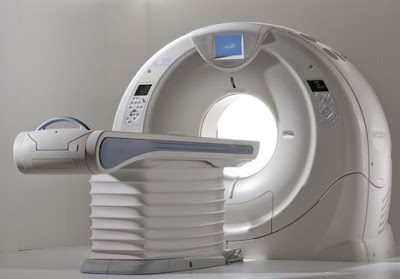
Toshiba's gargantuan dynamic volume system called AquilionONE, the device that features a coverage area of 320 detectors rows in addition to a respectable 650 lb table capacity, has now been installed at Beth Israel Deaconess Medical Center in Boston, according to a press release obtained by Medgadget. This is the third install of this device in the US. We first covered AquilionONE CT back in November 2007, when it was first unveiled at the RSNA 2007 conference. So when two days ago we were all excited about high demand for 256-slice CT scanners, we should have have kept a more proper perspective: the 320-slice system is also here to stay, albeit initially in smaller numbers. A representative for the company, tells Medgadget: "The Aquilion ONE has a coverage area of 320 detector rows, can capture actual organ movement (like blow flowing through the heart) and can image an entire organ in one gantry rotation. Additionally, the Aquilion ONE can capture the heart in one heart beat."

From the press release:
As a testament to the growing demand to improve patient care while reducing healthcare costs, Toshiba America Medical Systems, Inc. has installed the Aquilion ONE™ dynamic volume CT system at Beth Israel Deaconess Medical Center, a teaching hospital of Harvard Medical School in Boston.


"In one of the country's leading medical teaching hospitals, we hope the Aquilion ONE's ability to image an entire organ and show function for the first time will mean faster, more accurate diagnosis, better patient outcomes and ultimately lower healthcare costs for our patients," explained Dr. Vassilios D. Raptopoulos, interim radiologist-in-chief, Department of Radiology and director, CT services, Beth Israel Deaconess Medical Center. "We are grateful to be one of the first teaching hospitals in the United States using this advanced technology."


Toshiba's Aquilion ONE dynamic volume CT system utilizes 320 ultra-high resolution detector rows (0.5 mm in width) to image an entire organ in a single gantry rotation. The result is unparalleled in diagnostic imaging today and produces a 4D clinical video showing up to 16 cm of anatomical coverage, enough to capture the entire brain or heart, and show its movement such as blood flow.


"The Aquilion ONE has the potential to provide a single, comprehensive exam that can replace a variety of duplicative and invasive procedures," added Dr. Raptopoulos. "Its versatility and ability to diagnose disease fast will be used within our radiology department to detect and treat life-threatening conditions, including cancer, heart disease, stroke and other neurovascular conditions."


Espectacular!! Vejam no link mais imagens.

Link:

http://www.medical.toshiba.com/products/ct/DynamicVolume/ClinicalCardiac01.aspx

Reacção ao Gadolínio

Pois é...durante muito tempo apresentado como perfeito, sem história de reacções adversas, o Gadolínio revela agora as primeiras limitações.
Só agora, depois de alguns anos de práctica, começam a surgir os primeiros estudos de reacções adversas ao Gadolínio.
A Fibrose Sistémica Nefrogénica está a ser estudada como uma dessas recções que podem eventualmente estar relacionadas com a administração do Gadolínio.
Fica aqui um artigo interessante nesta matéria.
Link:

Nova Revista Científica Portuguesa


A ESTeS Lisboa lançou ontem a sua revista científica - Saúde & Tecnologia
Ainda só apresentada no site da ESTeSL, deve estar para breve a disponibilização da 1ª edição.
Como grande referência nacional no ensino das Tecnologias da Saúde, já justificava uma publicação deste tipo...agora é produzir artigos para mandar para lá...hehe


Artis Zee - Angiografia com imagens 3D


Lançada recentemente pela Siemens, esta máquina traz aquelas nossas imagens espectaculares 3D da TC para a sala de Angiografia, em tempo real...espectacular!!!


At the ongoing RSNA 2007 conference, Siemens has introduces a new family for interventional fluoroscopy imaging, a group of devices featuring multi-axis capabilities based on robotic technology. The first device in this class is Artis zeego, a system currently awaiting 510(k) approval by the FDA.


From the product brochure:

Artis Artis zeego is a multi-axis, angiographic system for interventional procedures. It has more freedom of positioning to accommodate nearly all projections. Fluoroscopy can be performed easily on the patient from head to toe. With a flexible isocenter that enables the physician to adjust the exam table to the most comfortable working position, operation is easy and precise. The syngo DynaCT application has been expanded and can be used even more flexibly for 3D reconstruction, because a larger volume is covered, expanding the view of the patient's anatomy. Not only are individual parts of the body imaged, but the entire abdomen. Images are acquired in landscape or portrait mode. In addition, Artis zeego's flexible park positions make it ideal for hybrid rooms. The very first Artis zeego system in the world is located at the Institute for Clinical Radiology of the University of Munich.


The Artis zee family portfolio also features dedicated C-arm systems for interventional cardiology and electrophysiology In this special field of intervention, excellent image quality is key. Imaging a moving structure such as the heart has always been a challenge. Now, with the Artis zee imaging system, outstanding image quality is provided from 2D fluoroscopy and 3D imaging and even a 4D image application where the time phases of the heartbeat are taken into account. In 2D fluoroscopy, features like advanced temporal filtration use an intelligent motion detection algorithm. This technology separates moving from non-moving structures in real time to improve the clarity of therapeutic instruments. Intelligent noise reduction enables high image quality during live fluoroscopy and acquisition by significantly reducing quantum noise without an increase in dose.


Stent meshes can be difficult to see, especially in obese patients or when steep angulations are used. But IC Stent* uses the balloon markers of the deployment balloon as reference points to shift and match images. Those images are then integrated to improve the signal-to-noise ratio to significantly enhance the visibility of stent meshes. For 3D applications, syngo IC3D helps to accurately measure lesions in the coronary arteries by using two projections. From these projections, a 3D model is generated so that a vessel can be rotated freely in space to precisely assess a lesion's diameter profile and the degree of stenosis. It also enables accurate measurement of lesion length to simplify appropriate stent selection.


The latest application, syngo DynaCT Cardiac, widens the 3D spectrum to 4D. By using rotational angiography and special reconstruction algorithms, syngo DynaCT Cardiac creates CT-like images of the beating heart right in the cath lab. During acquisition, it can use an ECG-triggered mode to collect image data to acquire only images from the same heart phase. With this feature it is now possible to reconstruct 4D images of the heart and its vessels in the cathlab.

Link:

http://www.medical.siemens.com/webapp/wcs/stores/servlet/PSGenericDisplay~q_catalogId~e_-1~a_catTree~e_100011,14209,1008807,1008805~a_langId~e_-1~a_pageId~e_95829~a_storeId~e_10001.htm

25 maio 2008

Forum das Tecnologias da Saúde


Para estrear este bloco de posts começo com este site, que admito que conheço há muito pouco tempo, mas que tem vindo a posicionar-se como um local de referência para os profissionais destas áreas.

Um site que aposta num intercâmbio de contacto, opiniões e ideias constante entre os profissionais, que apresenta um grupo de moderadores atento e que respondem rapidamente às questões colocadas, com muita informação para todos.

Também aceita Artigo e tem uma revista Online para a asua publicação.

Único em Portugal e, na minha opinião, muito importante.

Registem-se e contribuam para fazê-lo crescer e com ele as nossas profissões.

Link: http://www.tdtonline.org/forum/index.php

Jornadas de Radiologia Faro 2008


RM 3.0T GE

GE Healthcare Introduces New 3.0T MR Scanner


GE Healthcare announced it has received clearance from the U.S. Food and Drug Administration for the company's new 3.0T magnetic resonance (MR) scanner, the latest addition to GE's Signa family of MR systems.


The new Signa® MR750 3.0T was introduced at the International Society of Magnetic Resonance in Medicine in Toronto, May 3-9.


Equipped with the industry's most powerful gradients, easy-to-use workflow features and the company's advanced Thermal Management System, the Signa MR750 delivers up to 60 percent additional anatomical coverage and resolution unit per time. The system also allows for up to five times the imaging performance over previous generations, increasing the freedom for advanced application development, including: a routine liver exam in 15 minutes and a full breast exam in only two sequences.


"GE is committed to pushing the boundaries for MRI capabilities and we believe this new product will do that now and in the future," said Jim Davis, vice president and general manager of GE Healthcare's MR business. "We expect this product to be a robust and clinically capable MR system that will simplify MR exams without compromising quality or productivity."


The Signa MR750 features a newly designed RF Transmit system maximizing performance with a 17 percent gain in scanning efficiency. In addition, the system includes the GE-exclusive Optical RF Technology that adds up to 27 percent higher signal-to-noise ratio (SNR) over conventional, non-optical MR receivers by reducing electrical noise and increasing signal detection.


When combined with GE's use of high-density surface coils, the optical receive chain is a critical path for ensuring clear signal reception and data analysis. To ensure that the high-density approach will always be maintained, the Signa MR750 3.0T architecture is scalable to 128 channels of simultaneous data acquisition.


Revolutionizing imaging capabilities with new parallel imaging technique


In efforts to improve full-body imaging on 3.0T MR, GE Healthcare's Signa MR750 features a newly developed parallel imaging technique entitled ARC™, Auto Calibrating Reconstruction for Cartesian imaging. In addition to reducing specific absorption rate (SAR), the ARC technique also improves body imaging by allowing:

- Auto-calibration that helps avoid collecting external sensitivity map;

- Less sensitive to field-of-view (FOV) positioning with a tight FOV;

- Clinically practical reconstruction times for continuous scanning; and

- Workflow simplification that can be seamlessly integrated into sequences without the need for separate calibration scans.


As a result of this new imaging technique, the MR 750 also features new operational efficient advanced applications, including:


LAVA-IDEALTM


LAVA-IDEAL is a dual-echo acquisition technique that raises the bar on existing sequences to provide consistent, detailed, three-dimensional abdominal images in one breath-hold. By allowing the user to select the output image types-- in-phase, opposed-phase, water and fat-- LAVA-IDEAL has the ability to produce four image contrasts with only one scan. With this new 3.0T application, clinicians can now conduct a complete liver exam in 15 minutes. "LAVA-IDEAL is a robust sequence that offers 'fat only' and 'water only' images in addition to excellent in-phase and opposed-phase images in a single breath-hold. This sequence has quickly become a routine part of all our abdominal sequence protocols at 3.0T," said Dr. Elmar Merkle, professor of Radiology, Head of Body Magnetic Resonance Imaging and Medical Director of the Center for Advanced Magnetic Resonance Development at Duke University.


VIBRANT-IDEALTM


VIBRANT-IDEAL is a new application that allows for fat-free breast imaging with high spatio-temporal resolution. This application catches the shortest in- and out-of phase echoes to keep scan times comparable to single echo acquisitions even though twice the amount of data is collected. VIBRANT-IDEAL optimizes acquisition with a high signal-to-noise ratio (SNR) for acquiring high quality water and fat images. This capability lets the user prescribe thinner slices for high spatial resolution imaging.


PROPELLER 2.0


PROPELLER 2.0 enables strong performance in all neuro imaging planes with the implementation of the No Phase Wrap (NPW) technique. NPW allows virtually ghost-artifact-free, motion-immune scans in sagittal, coronal, axial and oblique planes. Since this technique effectively deals with the aliasing artifact, PROPELLER 2.0 is now more robust performing small field-of-view (FOV) scans.


Simplification of user interface, patient experience and automated acquisition and processing


Driven by customer demands for improved clinical capabilities, the Signa MR750 has been designed around clinical workflow needs with a strong focus on improving productivity, With that in mind, the system also includes operational efficiencies such as the newly designed detachable patient table and the first ever in-room operator console.


In addition, The Signa MR750 features automated acquisition based on the concept of touch and go protocols. The aspect of the system allows for more focused attention on the patient and consistent protocols and processing across operating technicians. Together, these features reduce setup time in the scanner room by up to 71 percent and additionally, the newly designed user interface reduces the number of steps by as much as 68 percent.


"MR users told us that they want to be able to provide the most advanced clinical applications to serve their communities, such as a complete liver exam in a 15-minute slot, routine fMRI with shorter paradigms and greater activation and a complete breast exam in only two sequences, but they need to be able to reproduce these exams very quickly and easily - every time," said David Handler, general manager of global MR marketing. "The Signa MR750 puts the most powerful MR technology at their fingertips to use in a very simple way so they can focus on patients."


Links: Steve Myers Cartoons

Os Links aqui do Blog vão sendo também aqui vistos mais ao pormenor, um a um.

Como primeiro link fica o site Innervisions - Fine Art X-ray Photography, de Steven Myers.


Trabalho excelente de um Técnico de Radiologia que se dedica à criação de imagens excelentes de radiografias de Flores.


No site mostra-nos as suas imagens preferidas assim como disponibiliza calendários e impressões a cores.

Definição do autor: Basic old school X-ray Photography.

Quem sabe mais uma opção de carreira? Vale a pena visitar.

Link: Steve Myers Cartoons

24 maio 2008

Apresentação das Jornadas do HPV

Os Diferentes Padrões da Doença Pulmonar em TC

Resumo:
Os contínuos desenvolvimentos da Radiologia moderna e aperfeiçoamento dos equipamentos de Tomografia Computorizada reforçam o estabelecimento desta técnica como gold standard para o estudo pormenorizado da Patologia Pulmonar.
O estudo da patologia pulmonar pela análise dos padrões de imagem que dela resultam é um método há muito instituído e globalmente aceite como o mais eficaz para o rápido estabelecimento da necessária correlação causa/efeito entre a patologia e a imagem que dela resulta.
O papel do Técnico de Radiologia, como primeiro avaliador da imagem adquirida, assume principal importância na identificação dos diferentes padrões resultantes e na necessária adaptação dos protocolos de aquisição para que possa fornecer ao Radiologista um estudo de grande detalhe da morfologia do Parênquima Pulmonar, que leve ao diagnóstico final.

Apresentado nas IIas Jornadas Técnicas de Radiologia do Centro Hospitalar Norte - Hospital Pulido Valente a 10 de Maio de 2008