Etiquetas
08 agosto 2008
05 agosto 2008
Processo de Bolonha - Parecer final
Afinal vai tudo ficar mesmo assim como está...uns licenciados de 3 anos, outros de 4.
Cabe às diferentes escolas decidir se fazem Licenciaturas de 3 ou 4 anos, sendo que, caso se decidam por 4 anos, o estágio profissional terá obrigatóriamente de estar incluido.
Uma decisão que acaba por agradar a todos...hehe
...a mim não...é pena que não nos consigamos entender porque acho que os cursos deveriam ser semelhantes, independentemente da Escola onde são leccionados...mas isso nunca acontece, não é?
As diferenças depois veêm-se cá fora...não vale de nada, mas dá para notar bem.
04 agosto 2008
É esperar que há-de chegar cá...
Resolution 208, Fairness in Medical Imaging Interpretation, is to come before the AMA's House of Delegates shortly, and it is expected to pass. It is introduced by some of our very good friends:
American Society of Neuroimaging
American Association of Neurological Surgeons
Congress of Neurological Surgeons
American Medical Group Association
American College of Cardiology
American College of Gastroenterology
American Gastroenterological Association
American Society for Gastrointestinal Endoscopy
(Associações de várias especialidades unidas por uma causa? Qual $erá a razão?)
25 julho 2008
HPP quer comprar SAMS

A CGD propôs a aquisição de 51% do SAMS ao sindicato dos bancários, que está aberto a negociar, avançou ontem o Expresso Online. O negócio inclui o Hospital dos Olivais e o Centro do Bairro Azul.
A CGD propôs a aquisição de 51% do SAMS ao sindicato dos bancários, que está aberto a negociar, avançou ontem o Expresso Online. O negócio inclui o Hospital dos Olivais e o Centro do Bairro Azul.A Hospitais Privados de Portugal, o braço do grupo da Caixa Geral de Depósitos para a área da saúde, quer entrar no capital dos SAMS. Segundo avança o Expresso Online, o acordo prevê a possibilidade de a HPP ficar com posições maioritárias nas instalações do hospital e postos médicos detidos pelos SAMS, assumindo ao mesmo tempo o controlo da gestão dos mesmos. Em contrapartida, os bancários filiados no sindicato poderão passar a ir a todos os hospitais da HPP, no caso de o acordo se consumar.Contactada pelo “Diário Económico”, que avança a mesma notícia na edição de hoje, a empresa não fez comentários, mas de acordo com as informações recolhidas pelo jornal, já foi entregue uma carta de intenções à administração dos SAMS, na qual se explica que o objectivo da parceria é que a HPP fique com 51% do capital da SAMS, incluindo o hospital dos Olivais e o centro médico do Bairro Azul, em Lisboa.O presidente do Sindicato dos Bancários do Sul e Ilhas (SBSI), Delmiro Carreira, adiantou ao DE que, por enquanto, apenas existe “uma declaração de intenções” por parte da HPP para negociar uma parceria. “O SBSI respondeu que estava disposto a negociar e agora vão começar as negociações que poderão dar origem a um protocolo e a um eventual negócio”, acrescentou Delmiro Carreira dizendo que "ainda não se falou de valores".
24 julho 2008
Novo Drug Eluting da Abbott
FDA Approves Abbott's Everolimus Eluting Stent

In May 2008, Abbott presented two-year data from the SPIRIT III trial demonstrating that XIENCE V continues to deliver positive clinical benefits for patients. At two years, the XIENCE V demonstrated the following key results:
- A 45 percent reduction in the risk of major adverse cardiac events (MACE) compared to TAXUS (7.3 percent for XIENCE V vs. 12.8 percent for TAXUS, p-value=0.004)*. MACE is an important composite clinical measure of safety and efficacy outcomes for patients, defined as cardiac death, heart attack (MI) or ischemia-driven target lesion revascularization (TLR, repeat procedures driven by lack of blood supply).
- A 32 percent reduction in the risk of TVF compared to TAXUS (10.7 percent for XIENCE V vs. 15.4 percent for TAXUS, p-value=0.04)*.
- Low rates of stent thrombosis between one and two years, defined as very late stent thrombosis, per Academic Research Consortium (ARC) definition of definite/probable stent thrombosis (0.3 percent for XIENCE V and 1.0 percent for TAXUS) and per the SPIRIT III protocol (0.2 percent for XIENCE V and 1.0 percent for TAXUS). The ARC definition of late stent thrombosis was developed to eliminate variability in the definitions across various drug eluting stent trials.
For more thought about the business behind the device, check out Jacob Goldstein's post over at the WSJ's Health Blog: Abbott Gets to Join Troubled Stent Market ...
Press release with video: FDA Approves Abbott's XIENCE™ V Drug Eluting Stent ...
Product page: XIENCE V ...
Link: www.medgadget.com
17 julho 2008
TC Crânio em doentes com AIT
NICE, France -- June 10, 2008 -- In the early phase after the onset of stroke-like neurological symptoms, cranial computer tomography (CCT) can neither depict nor rule out strokes in a reliable manner, according to the findings of a study presented here at the 18th Meeting of the European Neurological Society (ENS).
Transient ischaemic attacks (TIA) are defined as neurological symptoms lasting less than 24 hours without evidence of infarction. However, the risk for developing major strokes is increased in these patients and therefore necessitates immediate diagnostic work-up. Usually CCT is initially performed with the aim of ruling out other aetiologies such as haemorrhage, tumours, and major stroke.
"In order to justify exposure to radiation, the CCT should be sensitive enough to depict the underlying cause of symptoms," said principal investigator Stephan Ulmer, MD, Institute of Neuroradiology, University Hospital of Schleswig-Holstein, Kiel, Germany, in a poster presentation on June 9. "We assessed whether CCT scans are a sufficient imaging modality for monitoring TIA and whether the defined time window is applicable."
The researchers reviewed all CCT scans that were requested in patients with transient neurological symptoms at their clinics in a 1-year period. All studies that were requested for patients presenting with TIA were further analysed. Computed tomography scans (multirow-detector scanners), emergency notes, charts, and other performed imaging modalities such as magnetic resonance imaging were reviewed.
Patients that turned out to have experienced seizures with Todd paralysis rather than a TIA or other conditions that would require emergency imaging were excluded from the study.
Among 3,580 patients who underwent CCTs in the study period, 350 had been assessed due to transient neurological symptoms and were included in the study. 148 of these patients had experienced symptoms for less than 1 hour.
In contrast to the common recommendations, haemorrhage did not seem to be a matter of concern in this patient group. "There was not a single case of a haemorrhage when symptoms were confined to 1 hour," Dr. Ulmer emphasised. Only patients with symptoms beyond this time window were diagnosed with haemorrhagic strokes.
"We think that CCT is not a sufficient imaging method in patients whose symptoms resolve within 1 hour," Dr. Ulmer concluded. "Also, there might be a need to come up with a new definition of the TIA in terms of the defined time window."
He noted that these findings need to be confirmed on a larger scale, therefore a multicentre trial is being planned by the investigators.
[Presentation title: Is a Cranial Computer Tomography (CCT) Indicated in All Patients Suffering From Transient Ischemic Attack (TIA)? Abstract P350]
12 julho 2008
12 junho 2008
Revisão de doses CR e CT
The finding that unchecked CR doses in digital systems can escalate above required levels (and accepted international standards) has already led to an update in legislation and official National Radiation Dose Levels (NRDLs) in Luxembourg. Dr. Alexandra Schreiner, from the country's Ministry of Health, said the risks of unnecessarily high radiation doses are highest for children.
"The increase in risk is proportional to the increase in effective dose received by the patient," she said. "As far as the dose is concerned, the ALARA (as low as reasonably achievable) principle should always be applied if we want to work in an ethically correct manner, while dose reference levels should also be respected as far as legislation is concerned."
A videographic comparison of chest, pelvis, and lumbar spine x-rays following the conversion of three Luxembourg hospitals from film-screen to digital radiography revealed notable increases in entrance surface dose levels. However, the researchers warned that such changes go undetected by observers, radiographers, and physicians.
"Whereas if the dose increases on a film-screen system, it is immediately apparent, as the film will have a higher optical density and will become darker," Schreiner said. "The major risk with CR is that an increase in dose may pass unnoticed since it does not decrease image quality. For this reason, it is important that the CR system has a detector dose indicator and that the user be educated to look at it regularly."
By using automated quality control software for constancy testing and implementing protocols for acceptance testing and dose optimization, Schreiner's team reduced radiation doses significantly in areas where they were elevated. The NRDL for chest radiation was lowered by 20%, by 40% for the pelvis, and by 20% for the lumbar spine AP.
After optimization, CR radiation not only fell back from the elevated levels but dropped below the original levels as measured on conventional film-screen machines.
European legislation had earlier recorded a dose reference level for pelvis x-rays of 10 mGy; whereas after the dose-optimization program was implemented, CR was able to run successfully at just 5.85 mGy.
On the basis of these findings, Schreiner's dose-optimization work was rolled out in all hospitals across Luxembourg, supporting their switch to CR.
In devising the optimization protocols, the team referenced several U.S. and European reports on acceptance testing, constancy testing, and evaluating diagnostic processes. Digital radiography performance appraisal procedures, as compiled by the King's Centre for the Assessment of Radiological Equipment (KCARE) in London, were deemed to be "very efficient."
However, regular CR evaluation is required to ensure continued optimal performance, Schreiner added. Acceptance testing should be performed upon receipt of a new system, or when major components are changed, especially if they can influence dose or image quality.
Thereafter, a yearly quality control check by a medical physicist is recommended, while a radiographer should implement a monthly constancy test. This, it was concluded, would "ensure the best image quality with as low a dose as is achievable, as well as the safety of the patient."
CT radiation risk
In a separate study presented at ECR, Dr. Koos Geleijns of Leiden University Medical Center in the Netherlands provided a counterargument to recent studies criticizing the risks of radiation exposure in some radiology exams, such as CT angiography. Geleijns criticized current approaches for radiation risk assessment for focusing on single risk factors, and therefore being "inaccurate."
Discussing the failure to take into account short-term complications (including mortality) for disease and treatment, he said that common protocols underestimate these risks while overestimating radiation risk.
Geleijns proposed instead the use of a series of multiple-decrement "life tables," representing age- and gender-related functions pertaining to mortality, which integrate all relevant characteristics and risk factors for specific patient populations.
This method was applied in the study to coronary artery disease (CAD) diagnosis and follow-up of endovascular abdominal aortic aneurysm (AAA) repair. Mortality from radiation exposure and excess mortality from diseases and complications were included. Long-term mortality related to radiation exposure was estimated using the BEIR VII risk model. All risks were expressed as a reduction of life expectancy.
Results revealed that under clinical conditions the acute risk of mortality from cardiac catheterization in coronary artery disease was small (at 0.1%), yet it substantially exceeds radiation risks from either CAD (5 mSv) or coronary CT angiography (15 mSv).
High disease-related mortality associated with AAA (6% per year) far exceeds the radiation risks from CT follow-up (18 mSv/year), and also substantially lowers estimates of radiation risk when compared to current evaluative methodologies.
"We used the excess relative risk model, and the radiation risk is expressed relative to the background cancer risks," Geleijns noted. "With regard to the cancer risks, we looked at several organ groups as defined by the BEIR VII report."
Based on the results, the researchers recommend a comprehensive risk assessment be performed by using multiple-decrement life tables that integrate long- and short-term excess mortality. The next step is "to also include in the models the benefit and diagnosis and following treatment," Geleijns said.
By Rob Skelding
AuntMinnie.com contributing writer
March 9, 2008
Sugestão de estudo para Portugal
Based on responses to IMV’s 2007 CT Census Survey of U.S. Hospitals and Nonhospitals.
As of IMV's 2007 census survey, 81% of all installed CT systems had multislice detector capability, 18% had single-slice spiral detectors, and 1% had other detector types, including nonspiral detectors, electron beam CT (EBCT), and dual-source CT.
Quem gere o PACS? Informática ou Radiologia?
The premise of the argument presented by Dr. Paul J. Chang on behalf of IT department ownership is that PACS technology has become a component of the entire hospital informatics enterprise rather than its own unique entity. In an increasing number of hospital infrastructures, dedicated networks for PACS are unnecessary. Thick-client workstations are facing obsolescence. Virtualization of mass storage is replacing more costly dedicated image storage systems.
Modern healthcare IT should be structured as a matrix, according to Chang, who straddles both worlds as vice chairman of radiology informatics and director of pathology informatics at the University of Chicago Pritzker School of Medicine.
"You don't want to destroy domain expertise, but rather leverage it better. A matrix organization enables the experience of one domain to be adopted by another domain," he contended.
Because PACS is the multimedia component of an electronic health record (EHR), the EHR must be optimized to support radiology workflow. Not only is this a complex undertaking, but it logically fits as the responsibility of the IT department -- as long as the IT department has a global vision and a progressive philosophy, Chang said.
Dr. David Channin, chief of imaging informatics at Northwestern Memorial Hospital and the Feinberg School of Medicine in Chicago, disagreed.
"Radiology has led informatics technology innovation in hospitals and will continue to be the source of informatics leadership in healthcare," Channin said. "Domain expertise must take precedence over IT expertise. Tools don't drive domain innovation."
"If controlled in a central manner, such as a matrix structure, the priorities of a radiology department will be subjected to control by an IT department juggling priorities representing multiple domains in a hospital," he said. "If you don't have budgetary control of your bucket of allocated capital dollars, you have lost control. Your critically needed PACS upgrade will be competing with acquisition of a new laser doodad for OR."
Radiology departments should wield the power they have as cash cows for hospitals, define their IT domain borders, provide access to them with standard interfaces, and demand autonomy, according to Channin. He recommended that radiology departments contract with IT departments for "commodity services" such as networks, virtual operating systems, and data storage.
"As progressive as an IT department may be, it doesn't care about the quality of information that radiologists need," Channin said. "Even if a new capital acquisition is approved in February, you may be told that you can't upgrade XYZ until the department synchronizes something else in August. It is imperative that radiology departments be able to control their own domain."
By Cynthia Keen
Estudo U.S.A.

08 junho 2008
Caso Clínico - Teratoma
Um Caso muito interessante e não muito frequente que resultou, mais que na apresentação do Caso Clínico, na realização de um trabalho de revisão que fiz para mim.
Nunca o cheguei a apresentar em lado nenhum, apresento-o agora aqui.
06 junho 2008
AquilionONE da Toshiba - 320!!!
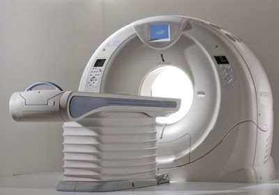
Toshiba's gargantuan dynamic volume system called AquilionONE, the device that features a coverage area of 320 detectors rows in addition to a respectable 650 lb table capacity, has now been installed at Beth Israel Deaconess Medical Center in Boston, according to a press release obtained by Medgadget. This is the third install of this device in the US. We first covered AquilionONE CT back in November 2007, when it was first unveiled at the RSNA 2007 conference. So when two days ago we were all excited about high demand for 256-slice CT scanners, we should have have kept a more proper perspective: the 320-slice system is also here to stay, albeit initially in smaller numbers. A representative for the company, tells Medgadget: "The Aquilion ONE has a coverage area of 320 detector rows, can capture actual organ movement (like blow flowing through the heart) and can image an entire organ in one gantry rotation. Additionally, the Aquilion ONE can capture the heart in one heart beat."
From the press release:
As a testament to the growing demand to improve patient care while reducing healthcare costs, Toshiba America Medical Systems, Inc. has installed the Aquilion ONE™ dynamic volume CT system at Beth Israel Deaconess Medical Center, a teaching hospital of Harvard Medical School in Boston.
"In one of the country's leading medical teaching hospitals, we hope the Aquilion ONE's ability to image an entire organ and show function for the first time will mean faster, more accurate diagnosis, better patient outcomes and ultimately lower healthcare costs for our patients," explained Dr. Vassilios D. Raptopoulos, interim radiologist-in-chief, Department of Radiology and director, CT services, Beth Israel Deaconess Medical Center. "We are grateful to be one of the first teaching hospitals in the United States using this advanced technology."
Toshiba's Aquilion ONE dynamic volume CT system utilizes 320 ultra-high resolution detector rows (0.5 mm in width) to image an entire organ in a single gantry rotation. The result is unparalleled in diagnostic imaging today and produces a 4D clinical video showing up to 16 cm of anatomical coverage, enough to capture the entire brain or heart, and show its movement such as blood flow.
"The Aquilion ONE has the potential to provide a single, comprehensive exam that can replace a variety of duplicative and invasive procedures," added Dr. Raptopoulos. "Its versatility and ability to diagnose disease fast will be used within our radiology department to detect and treat life-threatening conditions, including cancer, heart disease, stroke and other neurovascular conditions."

Espectacular!! Vejam no link mais imagens.
Link:
http://www.medical.toshiba.com/products/ct/DynamicVolume/ClinicalCardiac01.aspx
Reacção ao Gadolínio
Nova Revista Científica Portuguesa
