12 junho 2008

Revisão de doses CR e CT

New studies examine CR, CT radiation dose
By Rob Skelding
AuntMinnie.com contributing writer
March 9, 2008
VIENNA - A five-year study presented Sunday at the 2008 European Congress of Radiology (ECR) spotlighted the importance of x-ray dose optimization for computed radiography (CR) systems, particularly when converting to CR from conventional film-screen equipment.

The finding that unchecked CR doses in digital systems can escalate above required levels (and accepted international standards) has already led to an update in legislation and official National Radiation Dose Levels (NRDLs) in Luxembourg. Dr. Alexandra Schreiner, from the country's Ministry of Health, said the risks of unnecessarily high radiation doses are highest for children.

"The increase in risk is proportional to the increase in effective dose received by the patient," she said. "As far as the dose is concerned, the ALARA (as low as reasonably achievable) principle should always be applied if we want to work in an ethically correct manner, while dose reference levels should also be respected as far as legislation is concerned."

A videographic comparison of chest, pelvis, and lumbar spine x-rays following the conversion of three Luxembourg hospitals from film-screen to digital radiography revealed notable increases in entrance surface dose levels. However, the researchers warned that such changes go undetected by observers, radiographers, and physicians.

"Whereas if the dose increases on a film-screen system, it is immediately apparent, as the film will have a higher optical density and will become darker," Schreiner said. "The major risk with CR is that an increase in dose may pass unnoticed since it does not decrease image quality. For this reason, it is important that the CR system has a detector dose indicator and that the user be educated to look at it regularly."

By using automated quality control software for constancy testing and implementing protocols for acceptance testing and dose optimization, Schreiner's team reduced radiation doses significantly in areas where they were elevated. The NRDL for chest radiation was lowered by 20%, by 40% for the pelvis, and by 20% for the lumbar spine AP.

After optimization, CR radiation not only fell back from the elevated levels but dropped below the original levels as measured on conventional film-screen machines.

European legislation had earlier recorded a dose reference level for pelvis x-rays of 10 mGy; whereas after the dose-optimization program was implemented, CR was able to run successfully at just 5.85 mGy.

On the basis of these findings, Schreiner's dose-optimization work was rolled out in all hospitals across Luxembourg, supporting their switch to CR.

In devising the optimization protocols, the team referenced several U.S. and European reports on acceptance testing, constancy testing, and evaluating diagnostic processes. Digital radiography performance appraisal procedures, as compiled by the King's Centre for the Assessment of Radiological Equipment (KCARE) in London, were deemed to be "very efficient."

However, regular CR evaluation is required to ensure continued optimal performance, Schreiner added. Acceptance testing should be performed upon receipt of a new system, or when major components are changed, especially if they can influence dose or image quality.

Thereafter, a yearly quality control check by a medical physicist is recommended, while a radiographer should implement a monthly constancy test. This, it was concluded, would "ensure the best image quality with as low a dose as is achievable, as well as the safety of the patient."

CT radiation risk

In a separate study presented at ECR, Dr. Koos Geleijns of Leiden University Medical Center in the Netherlands provided a counterargument to recent studies criticizing the risks of radiation exposure in some radiology exams, such as CT angiography. Geleijns criticized current approaches for radiation risk assessment for focusing on single risk factors, and therefore being "inaccurate."

Discussing the failure to take into account short-term complications (including mortality) for disease and treatment, he said that common protocols underestimate these risks while overestimating radiation risk.

Geleijns proposed instead the use of a series of multiple-decrement "life tables," representing age- and gender-related functions pertaining to mortality, which integrate all relevant characteristics and risk factors for specific patient populations.

This method was applied in the study to coronary artery disease (CAD) diagnosis and follow-up of endovascular abdominal aortic aneurysm (AAA) repair. Mortality from radiation exposure and excess mortality from diseases and complications were included. Long-term mortality related to radiation exposure was estimated using the BEIR VII risk model. All risks were expressed as a reduction of life expectancy.

Results revealed that under clinical conditions the acute risk of mortality from cardiac catheterization in coronary artery disease was small (at 0.1%), yet it substantially exceeds radiation risks from either CAD (5 mSv) or coronary CT angiography (15 mSv).

High disease-related mortality associated with AAA (6% per year) far exceeds the radiation risks from CT follow-up (18 mSv/year), and also substantially lowers estimates of radiation risk when compared to current evaluative methodologies.

"We used the excess relative risk model, and the radiation risk is expressed relative to the background cancer risks," Geleijns noted. "With regard to the cancer risks, we looked at several organ groups as defined by the BEIR VII report."

Based on the results, the researchers recommend a comprehensive risk assessment be performed by using multiple-decrement life tables that integrate long- and short-term excess mortality. The next step is "to also include in the models the benefit and diagnosis and following treatment," Geleijns said.

By Rob Skelding
AuntMinnie.com contributing writer
March 9, 2008

Sugestão de estudo para Portugal

MarketStat #50: Single versus multislice detectors in CT installed base
March 31, 2008

Based on responses to IMV’s 2007 CT Census Survey of U.S. Hospitals and Nonhospitals.
As of IMV's 2007 census survey, 81% of all installed CT systems had multislice detector capability, 18% had single-slice spiral detectors, and 1% had other detector types, including nonspiral detectors, electron beam CT (EBCT), and dual-source CT.

Quem gere o PACS? Informática ou Radiologia?

Uma interessante discussão relativamente à atribuição da responsabilidade de gestão do sistema PACS. Deverá esta ser entregue aos serviços de Informática ou à Radiologia? Os argumentos dos dois lados desta discussão bem actual.
Who owns PACS -- Radiology or IT?
By Cynthia KeenJune 4, 2008
Should radiology or the IT department take responsibility for managing PACS in a hospital? It depends on the facility's corporate culture and the level of sophistication of the IT department, an animated "debate" at the 2008 Society for Imaging Informatics in Medicine (SIIM) meeting concluded.
In a typical client server environment data resides in a traditional, direct-attached storage model, randomly distributed across the enterprise. The result is silos of information that function sufficiently, but only when proximity and connectivity to the feeder systems is maintained. Healthcare institutions are forced to plan on ways to communicate patient administrative data, orders and results (i.e., laboratory, radiology, pharmacy and clinical doc) .

The premise of the argument presented by Dr. Paul J. Chang on behalf of IT department ownership is that PACS technology has become a component of the entire hospital informatics enterprise rather than its own unique entity. In an increasing number of hospital infrastructures, dedicated networks for PACS are unnecessary. Thick-client workstations are facing obsolescence. Virtualization of mass storage is replacing more costly dedicated image storage systems.

Modern healthcare IT should be structured as a matrix, according to Chang, who straddles both worlds as vice chairman of radiology informatics and director of pathology informatics at the University of Chicago Pritzker School of Medicine.
"You don't want to destroy domain expertise, but rather leverage it better. A matrix organization enables the experience of one domain to be adopted by another domain," he contended.

Because PACS is the multimedia component of an electronic health record (EHR), the EHR must be optimized to support radiology workflow. Not only is this a complex undertaking, but it logically fits as the responsibility of the IT department -- as long as the IT department has a global vision and a progressive philosophy, Chang said.

Dr. David Channin, chief of imaging informatics at Northwestern Memorial Hospital and the Feinberg School of Medicine in Chicago, disagreed.

"Radiology has led informatics technology innovation in hospitals and will continue to be the source of informatics leadership in healthcare," Channin said. "Domain expertise must take precedence over IT expertise. Tools don't drive domain innovation."

"If controlled in a central manner, such as a matrix structure, the priorities of a radiology department will be subjected to control by an IT department juggling priorities representing multiple domains in a hospital," he said. "If you don't have budgetary control of your bucket of allocated capital dollars, you have lost control. Your critically needed PACS upgrade will be competing with acquisition of a new laser doodad for OR."

Radiology departments should wield the power they have as cash cows for hospitals, define their IT domain borders, provide access to them with standard interfaces, and demand autonomy, according to Channin. He recommended that radiology departments contract with IT departments for "commodity services" such as networks, virtual operating systems, and data storage.

"As progressive as an IT department may be, it doesn't care about the quality of information that radiologists need," Channin said. "Even if a new capital acquisition is approved in February, you may be told that you can't upgrade XYZ until the department synchronizes something else in August. It is imperative that radiology departments be able to control their own domain."
AuntMinnie.com contributing writer
June 4, 2008

Estudo U.S.A.


A study has found that one in 20 patients have had the urge to kill their doctor. The survey questioned 800 Americans who were in pain, undergoing physical rehabilitation or seeking legal compensation for disability to find out their attitude towards their GP. The researchers from the University of Miami, Florida, found that just over 1 in 20, or five per cent, admitted feeling like they wanted to murder their physician.

08 junho 2008

Caso Clínico - Teratoma

Um Caso muito interessante e não muito frequente que resultou, mais que na apresentação do Caso Clínico, na realização de um trabalho de revisão que fiz para mim.


Nunca o cheguei a apresentar em lado nenhum, apresento-o agora aqui.

06 junho 2008

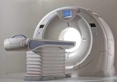
AquilionONE da Toshiba - 320!!!

Eu quero um destes...hehehehe


Toshiba's gargantuan dynamic volume system called AquilionONE, the device that features a coverage area of 320 detectors rows in addition to a respectable 650 lb table capacity, has now been installed at Beth Israel Deaconess Medical Center in Boston, according to a press release obtained by Medgadget. This is the third install of this device in the US. We first covered AquilionONE CT back in November 2007, when it was first unveiled at the RSNA 2007 conference. So when two days ago we were all excited about high demand for 256-slice CT scanners, we should have have kept a more proper perspective: the 320-slice system is also here to stay, albeit initially in smaller numbers. A representative for the company, tells Medgadget: "The Aquilion ONE has a coverage area of 320 detector rows, can capture actual organ movement (like blow flowing through the heart) and can image an entire organ in one gantry rotation. Additionally, the Aquilion ONE can capture the heart in one heart beat."

From the press release:
As a testament to the growing demand to improve patient care while reducing healthcare costs, Toshiba America Medical Systems, Inc. has installed the Aquilion ONE™ dynamic volume CT system at Beth Israel Deaconess Medical Center, a teaching hospital of Harvard Medical School in Boston.


"In one of the country's leading medical teaching hospitals, we hope the Aquilion ONE's ability to image an entire organ and show function for the first time will mean faster, more accurate diagnosis, better patient outcomes and ultimately lower healthcare costs for our patients," explained Dr. Vassilios D. Raptopoulos, interim radiologist-in-chief, Department of Radiology and director, CT services, Beth Israel Deaconess Medical Center. "We are grateful to be one of the first teaching hospitals in the United States using this advanced technology."


Toshiba's Aquilion ONE dynamic volume CT system utilizes 320 ultra-high resolution detector rows (0.5 mm in width) to image an entire organ in a single gantry rotation. The result is unparalleled in diagnostic imaging today and produces a 4D clinical video showing up to 16 cm of anatomical coverage, enough to capture the entire brain or heart, and show its movement such as blood flow.


"The Aquilion ONE has the potential to provide a single, comprehensive exam that can replace a variety of duplicative and invasive procedures," added Dr. Raptopoulos. "Its versatility and ability to diagnose disease fast will be used within our radiology department to detect and treat life-threatening conditions, including cancer, heart disease, stroke and other neurovascular conditions."


Espectacular!! Vejam no link mais imagens.

Link:

http://www.medical.toshiba.com/products/ct/DynamicVolume/ClinicalCardiac01.aspx

Reacção ao Gadolínio

Pois é...durante muito tempo apresentado como perfeito, sem história de reacções adversas, o Gadolínio revela agora as primeiras limitações.
Só agora, depois de alguns anos de práctica, começam a surgir os primeiros estudos de reacções adversas ao Gadolínio.
A Fibrose Sistémica Nefrogénica está a ser estudada como uma dessas recções que podem eventualmente estar relacionadas com a administração do Gadolínio.
Fica aqui um artigo interessante nesta matéria.
Link:

Nova Revista Científica Portuguesa


A ESTeS Lisboa lançou ontem a sua revista científica - Saúde & Tecnologia
Ainda só apresentada no site da ESTeSL, deve estar para breve a disponibilização da 1ª edição.
Como grande referência nacional no ensino das Tecnologias da Saúde, já justificava uma publicação deste tipo...agora é produzir artigos para mandar para lá...hehe


Artis Zee - Angiografia com imagens 3D


Lançada recentemente pela Siemens, esta máquina traz aquelas nossas imagens espectaculares 3D da TC para a sala de Angiografia, em tempo real...espectacular!!!


At the ongoing RSNA 2007 conference, Siemens has introduces a new family for interventional fluoroscopy imaging, a group of devices featuring multi-axis capabilities based on robotic technology. The first device in this class is Artis zeego, a system currently awaiting 510(k) approval by the FDA.


From the product brochure:

Artis Artis zeego is a multi-axis, angiographic system for interventional procedures. It has more freedom of positioning to accommodate nearly all projections. Fluoroscopy can be performed easily on the patient from head to toe. With a flexible isocenter that enables the physician to adjust the exam table to the most comfortable working position, operation is easy and precise. The syngo DynaCT application has been expanded and can be used even more flexibly for 3D reconstruction, because a larger volume is covered, expanding the view of the patient's anatomy. Not only are individual parts of the body imaged, but the entire abdomen. Images are acquired in landscape or portrait mode. In addition, Artis zeego's flexible park positions make it ideal for hybrid rooms. The very first Artis zeego system in the world is located at the Institute for Clinical Radiology of the University of Munich.


The Artis zee family portfolio also features dedicated C-arm systems for interventional cardiology and electrophysiology In this special field of intervention, excellent image quality is key. Imaging a moving structure such as the heart has always been a challenge. Now, with the Artis zee imaging system, outstanding image quality is provided from 2D fluoroscopy and 3D imaging and even a 4D image application where the time phases of the heartbeat are taken into account. In 2D fluoroscopy, features like advanced temporal filtration use an intelligent motion detection algorithm. This technology separates moving from non-moving structures in real time to improve the clarity of therapeutic instruments. Intelligent noise reduction enables high image quality during live fluoroscopy and acquisition by significantly reducing quantum noise without an increase in dose.


Stent meshes can be difficult to see, especially in obese patients or when steep angulations are used. But IC Stent* uses the balloon markers of the deployment balloon as reference points to shift and match images. Those images are then integrated to improve the signal-to-noise ratio to significantly enhance the visibility of stent meshes. For 3D applications, syngo IC3D helps to accurately measure lesions in the coronary arteries by using two projections. From these projections, a 3D model is generated so that a vessel can be rotated freely in space to precisely assess a lesion's diameter profile and the degree of stenosis. It also enables accurate measurement of lesion length to simplify appropriate stent selection.


The latest application, syngo DynaCT Cardiac, widens the 3D spectrum to 4D. By using rotational angiography and special reconstruction algorithms, syngo DynaCT Cardiac creates CT-like images of the beating heart right in the cath lab. During acquisition, it can use an ECG-triggered mode to collect image data to acquire only images from the same heart phase. With this feature it is now possible to reconstruct 4D images of the heart and its vessels in the cathlab.

Link:

http://www.medical.siemens.com/webapp/wcs/stores/servlet/PSGenericDisplay~q_catalogId~e_-1~a_catTree~e_100011,14209,1008807,1008805~a_langId~e_-1~a_pageId~e_95829~a_storeId~e_10001.htm

25 maio 2008

Forum das Tecnologias da Saúde


Para estrear este bloco de posts começo com este site, que admito que conheço há muito pouco tempo, mas que tem vindo a posicionar-se como um local de referência para os profissionais destas áreas.

Um site que aposta num intercâmbio de contacto, opiniões e ideias constante entre os profissionais, que apresenta um grupo de moderadores atento e que respondem rapidamente às questões colocadas, com muita informação para todos.

Também aceita Artigo e tem uma revista Online para a asua publicação.

Único em Portugal e, na minha opinião, muito importante.

Registem-se e contribuam para fazê-lo crescer e com ele as nossas profissões.

Link: http://www.tdtonline.org/forum/index.php

Jornadas de Radiologia Faro 2008


RM 3.0T GE

GE Healthcare Introduces New 3.0T MR Scanner


GE Healthcare announced it has received clearance from the U.S. Food and Drug Administration for the company's new 3.0T magnetic resonance (MR) scanner, the latest addition to GE's Signa family of MR systems.


The new Signa® MR750 3.0T was introduced at the International Society of Magnetic Resonance in Medicine in Toronto, May 3-9.


Equipped with the industry's most powerful gradients, easy-to-use workflow features and the company's advanced Thermal Management System, the Signa MR750 delivers up to 60 percent additional anatomical coverage and resolution unit per time. The system also allows for up to five times the imaging performance over previous generations, increasing the freedom for advanced application development, including: a routine liver exam in 15 minutes and a full breast exam in only two sequences.


"GE is committed to pushing the boundaries for MRI capabilities and we believe this new product will do that now and in the future," said Jim Davis, vice president and general manager of GE Healthcare's MR business. "We expect this product to be a robust and clinically capable MR system that will simplify MR exams without compromising quality or productivity."


The Signa MR750 features a newly designed RF Transmit system maximizing performance with a 17 percent gain in scanning efficiency. In addition, the system includes the GE-exclusive Optical RF Technology that adds up to 27 percent higher signal-to-noise ratio (SNR) over conventional, non-optical MR receivers by reducing electrical noise and increasing signal detection.


When combined with GE's use of high-density surface coils, the optical receive chain is a critical path for ensuring clear signal reception and data analysis. To ensure that the high-density approach will always be maintained, the Signa MR750 3.0T architecture is scalable to 128 channels of simultaneous data acquisition.


Revolutionizing imaging capabilities with new parallel imaging technique


In efforts to improve full-body imaging on 3.0T MR, GE Healthcare's Signa MR750 features a newly developed parallel imaging technique entitled ARC™, Auto Calibrating Reconstruction for Cartesian imaging. In addition to reducing specific absorption rate (SAR), the ARC technique also improves body imaging by allowing:

- Auto-calibration that helps avoid collecting external sensitivity map;

- Less sensitive to field-of-view (FOV) positioning with a tight FOV;

- Clinically practical reconstruction times for continuous scanning; and

- Workflow simplification that can be seamlessly integrated into sequences without the need for separate calibration scans.


As a result of this new imaging technique, the MR 750 also features new operational efficient advanced applications, including:


LAVA-IDEALTM


LAVA-IDEAL is a dual-echo acquisition technique that raises the bar on existing sequences to provide consistent, detailed, three-dimensional abdominal images in one breath-hold. By allowing the user to select the output image types-- in-phase, opposed-phase, water and fat-- LAVA-IDEAL has the ability to produce four image contrasts with only one scan. With this new 3.0T application, clinicians can now conduct a complete liver exam in 15 minutes. "LAVA-IDEAL is a robust sequence that offers 'fat only' and 'water only' images in addition to excellent in-phase and opposed-phase images in a single breath-hold. This sequence has quickly become a routine part of all our abdominal sequence protocols at 3.0T," said Dr. Elmar Merkle, professor of Radiology, Head of Body Magnetic Resonance Imaging and Medical Director of the Center for Advanced Magnetic Resonance Development at Duke University.


VIBRANT-IDEALTM


VIBRANT-IDEAL is a new application that allows for fat-free breast imaging with high spatio-temporal resolution. This application catches the shortest in- and out-of phase echoes to keep scan times comparable to single echo acquisitions even though twice the amount of data is collected. VIBRANT-IDEAL optimizes acquisition with a high signal-to-noise ratio (SNR) for acquiring high quality water and fat images. This capability lets the user prescribe thinner slices for high spatial resolution imaging.


PROPELLER 2.0


PROPELLER 2.0 enables strong performance in all neuro imaging planes with the implementation of the No Phase Wrap (NPW) technique. NPW allows virtually ghost-artifact-free, motion-immune scans in sagittal, coronal, axial and oblique planes. Since this technique effectively deals with the aliasing artifact, PROPELLER 2.0 is now more robust performing small field-of-view (FOV) scans.


Simplification of user interface, patient experience and automated acquisition and processing


Driven by customer demands for improved clinical capabilities, the Signa MR750 has been designed around clinical workflow needs with a strong focus on improving productivity, With that in mind, the system also includes operational efficiencies such as the newly designed detachable patient table and the first ever in-room operator console.


In addition, The Signa MR750 features automated acquisition based on the concept of touch and go protocols. The aspect of the system allows for more focused attention on the patient and consistent protocols and processing across operating technicians. Together, these features reduce setup time in the scanner room by up to 71 percent and additionally, the newly designed user interface reduces the number of steps by as much as 68 percent.


"MR users told us that they want to be able to provide the most advanced clinical applications to serve their communities, such as a complete liver exam in a 15-minute slot, routine fMRI with shorter paradigms and greater activation and a complete breast exam in only two sequences, but they need to be able to reproduce these exams very quickly and easily - every time," said David Handler, general manager of global MR marketing. "The Signa MR750 puts the most powerful MR technology at their fingertips to use in a very simple way so they can focus on patients."


Links: Steve Myers Cartoons

Os Links aqui do Blog vão sendo também aqui vistos mais ao pormenor, um a um.

Como primeiro link fica o site Innervisions - Fine Art X-ray Photography, de Steven Myers.


Trabalho excelente de um Técnico de Radiologia que se dedica à criação de imagens excelentes de radiografias de Flores.


No site mostra-nos as suas imagens preferidas assim como disponibiliza calendários e impressões a cores.

Definição do autor: Basic old school X-ray Photography.

Quem sabe mais uma opção de carreira? Vale a pena visitar.

Link: Steve Myers Cartoons

24 maio 2008

Apresentação das Jornadas do HPV

Os Diferentes Padrões da Doença Pulmonar em TC

Resumo:
Os contínuos desenvolvimentos da Radiologia moderna e aperfeiçoamento dos equipamentos de Tomografia Computorizada reforçam o estabelecimento desta técnica como gold standard para o estudo pormenorizado da Patologia Pulmonar.
O estudo da patologia pulmonar pela análise dos padrões de imagem que dela resultam é um método há muito instituído e globalmente aceite como o mais eficaz para o rápido estabelecimento da necessária correlação causa/efeito entre a patologia e a imagem que dela resulta.
O papel do Técnico de Radiologia, como primeiro avaliador da imagem adquirida, assume principal importância na identificação dos diferentes padrões resultantes e na necessária adaptação dos protocolos de aquisição para que possa fornecer ao Radiologista um estudo de grande detalhe da morfologia do Parênquima Pulmonar, que leve ao diagnóstico final.

Apresentado nas IIas Jornadas Técnicas de Radiologia do Centro Hospitalar Norte - Hospital Pulido Valente a 10 de Maio de 2008