
Outro projecto de voluntariado internacional.
Link: http://www.rad-aid.org/


Interesting snippet out of the UK. A research group of forensic pathologists and radiologists at Leicester University claims to have developed a minimally invasive autopsy method.Professor Guy Rutty, Chief Forensic Pathologist to the East Midlands Forensic Pathology Unit at University of Leicester, explains:“In collaboration with the radiology team, lead by Professor Bruno Morgan, we have successfully developed a quick and simple technique of ‘minimally invasive targeted coronary angiography’ where we inject contrast into the body of a deceased person through a small incision in the neck and then perform a full body CT scan. Using this method we are able to determine the cause of death in up to 80% of cases (in the series analysed to date).


Merge real-time ultrasound with previously acquired CT, MR or ultrasound images.

- Low rates of stent thrombosis between one and two years, defined as very late stent thrombosis, per Academic Research Consortium (ARC) definition of definite/probable stent thrombosis (0.3 percent for XIENCE V and 1.0 percent for TAXUS) and per the SPIRIT III protocol (0.2 percent for XIENCE V and 1.0 percent for TAXUS). The ARC definition of late stent thrombosis was developed to eliminate variability in the definitions across various drug eluting stent trials.
For more thought about the business behind the device, check out Jacob Goldstein's post over at the WSJ's Health Blog: Abbott Gets to Join Troubled Stent Market ...
Press release with video: FDA Approves Abbott's XIENCE™ V Drug Eluting Stent ...
Product page: XIENCE V ...
Link: www.medgadget.com
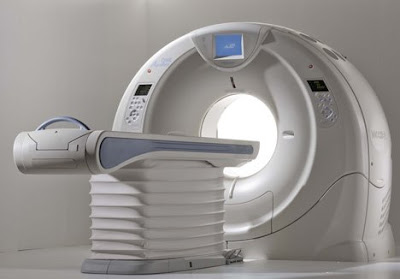
Toshiba's gargantuan dynamic volume system called AquilionONE, the device that features a coverage area of 320 detectors rows in addition to a respectable 650 lb table capacity, has now been installed at Beth Israel Deaconess Medical Center in Boston, according to a press release obtained by Medgadget. This is the third install of this device in the US. We first covered AquilionONE CT back in November 2007, when it was first unveiled at the RSNA 2007 conference. So when two days ago we were all excited about high demand for 256-slice CT scanners, we should have have kept a more proper perspective: the 320-slice system is also here to stay, albeit initially in smaller numbers. A representative for the company, tells Medgadget: "The Aquilion ONE has a coverage area of 320 detector rows, can capture actual organ movement (like blow flowing through the heart) and can image an entire organ in one gantry rotation. Additionally, the Aquilion ONE can capture the heart in one heart beat."
From the press release:
As a testament to the growing demand to improve patient care while reducing healthcare costs, Toshiba America Medical Systems, Inc. has installed the Aquilion ONE™ dynamic volume CT system at Beth Israel Deaconess Medical Center, a teaching hospital of Harvard Medical School in Boston.
"In one of the country's leading medical teaching hospitals, we hope the Aquilion ONE's ability to image an entire organ and show function for the first time will mean faster, more accurate diagnosis, better patient outcomes and ultimately lower healthcare costs for our patients," explained Dr. Vassilios D. Raptopoulos, interim radiologist-in-chief, Department of Radiology and director, CT services, Beth Israel Deaconess Medical Center. "We are grateful to be one of the first teaching hospitals in the United States using this advanced technology."
Toshiba's Aquilion ONE dynamic volume CT system utilizes 320 ultra-high resolution detector rows (0.5 mm in width) to image an entire organ in a single gantry rotation. The result is unparalleled in diagnostic imaging today and produces a 4D clinical video showing up to 16 cm of anatomical coverage, enough to capture the entire brain or heart, and show its movement such as blood flow.
"The Aquilion ONE has the potential to provide a single, comprehensive exam that can replace a variety of duplicative and invasive procedures," added Dr. Raptopoulos. "Its versatility and ability to diagnose disease fast will be used within our radiology department to detect and treat life-threatening conditions, including cancer, heart disease, stroke and other neurovascular conditions."

Link:
http://www.medical.toshiba.com/products/ct/DynamicVolume/ClinicalCardiac01.aspx

At the ongoing RSNA 2007 conference, Siemens has introduces a new family for interventional fluoroscopy imaging, a group of devices featuring multi-axis capabilities based on robotic technology. The first device in this class is Artis zeego, a system currently awaiting 510(k) approval by the FDA.
From the product brochure:
Artis Artis zeego is a multi-axis, angiographic system for interventional procedures. It has more freedom of positioning to accommodate nearly all projections. Fluoroscopy can be performed easily on the patient from head to toe. With a flexible isocenter that enables the physician to adjust the exam table to the most comfortable working position, operation is easy and precise. The syngo DynaCT application has been expanded and can be used even more flexibly for 3D reconstruction, because a larger volume is covered, expanding the view of the patient's anatomy. Not only are individual parts of the body imaged, but the entire abdomen. Images are acquired in landscape or portrait mode. In addition, Artis zeego's flexible park positions make it ideal for hybrid rooms. The very first Artis zeego system in the world is located at the Institute for Clinical Radiology of the University of Munich.
The Artis zee family portfolio also features dedicated C-arm systems for interventional cardiology and electrophysiology In this special field of intervention, excellent image quality is key. Imaging a moving structure such as the heart has always been a challenge. Now, with the Artis zee imaging system, outstanding image quality is provided from 2D fluoroscopy and 3D imaging and even a 4D image application where the time phases of the heartbeat are taken into account. In 2D fluoroscopy, features like advanced temporal filtration use an intelligent motion detection algorithm. This technology separates moving from non-moving structures in real time to improve the clarity of therapeutic instruments. Intelligent noise reduction enables high image quality during live fluoroscopy and acquisition by significantly reducing quantum noise without an increase in dose.
Stent meshes can be difficult to see, especially in obese patients or when steep angulations are used. But IC Stent* uses the balloon markers of the deployment balloon as reference points to shift and match images. Those images are then integrated to improve the signal-to-noise ratio to significantly enhance the visibility of stent meshes. For 3D applications, syngo IC3D helps to accurately measure lesions in the coronary arteries by using two projections. From these projections, a 3D model is generated so that a vessel can be rotated freely in space to precisely assess a lesion's diameter profile and the degree of stenosis. It also enables accurate measurement of lesion length to simplify appropriate stent selection.
The latest application, syngo DynaCT Cardiac, widens the 3D spectrum to 4D. By using rotational angiography and special reconstruction algorithms, syngo DynaCT Cardiac creates CT-like images of the beating heart right in the cath lab. During acquisition, it can use an ECG-triggered mode to collect image data to acquire only images from the same heart phase. With this feature it is now possible to reconstruct 4D images of the heart and its vessels in the cathlab.
Link:


